60 Strathallen Avenue, Northbridge NSW
Monday to Friday - 9am to 5pm
Our Technology
Our eye clinic is equipped with state-of-the-art equipment sourced from around the world, each operated by highly trained and experienced orthoptists (ophthalmic technicians). We use only the latest and best technology to ensure that all our patients receive the highest standard of care.
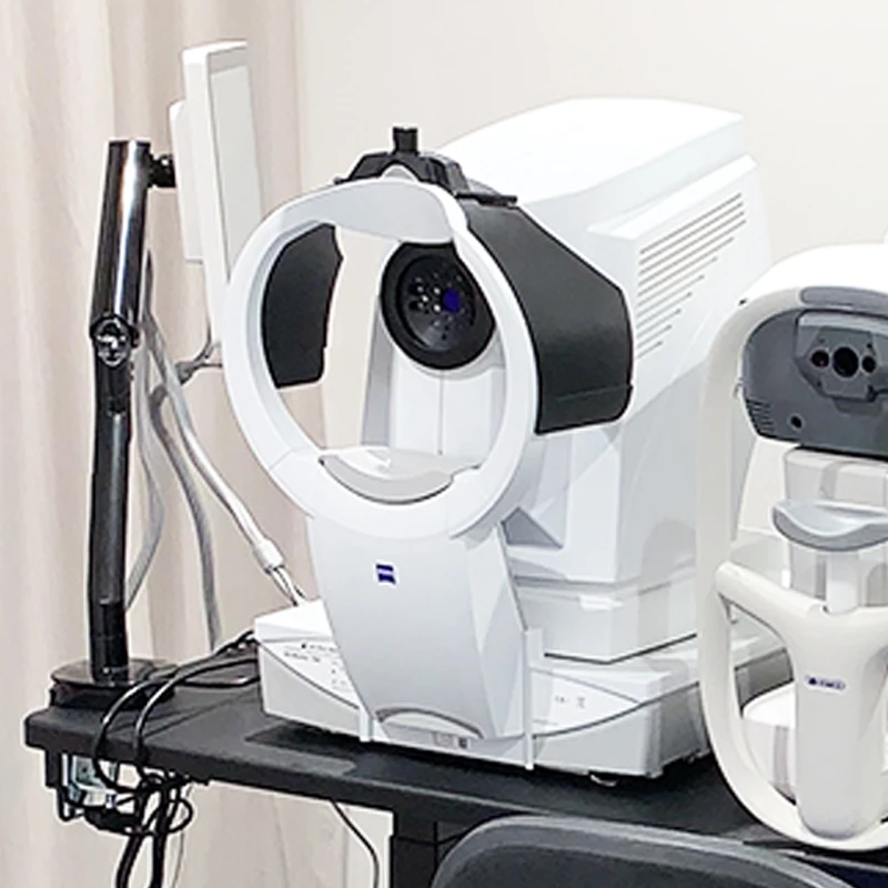
Ocular Biometry with Zeiss IOLMaster
Used for complex eye measurements
The IOL Master is a non-contact optical coherence biometer which allows for complex eye length and surface curvature measurements to be gathered without the use of anesthesia, and minimising the potential for contamination.
During cataract surgery, the cataract is removed and a prosthetic intraocular lens (IOL) implanted in its place. Highly detailed measurements of the eye are taken before surgery which allow our surgeons to calculate the correct type and power of IOL to implant.
Using technology such as partial coherence interferometry, the IOL Master provides consistently precise results, which are required in complex calculations made by our surgeons before cataract surgery.
An internal algorithm allows for an accurate calculation of the distance to the vitreoretinal interface (where the vitreous gel meets the retina at the back of the eye), which is calibrated against a high resolution biometric system, and allows the accurate selection of an IOL to suit each individual patient.
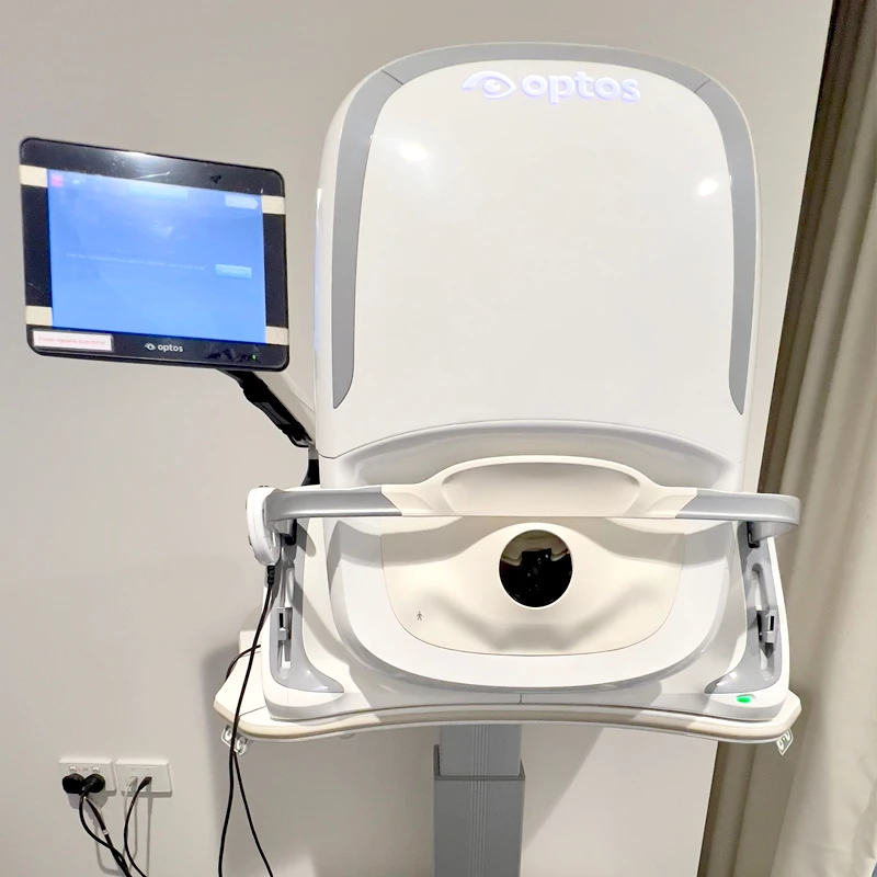
Retinal Imaging with Optos
Captures specific details of the retina and optic nerve
The Heidelberg HRA uses confocal scanning laser technology to allow our ophthalmologists to use advanced imaging modes as well as traditional methods to evaluate the retina and optic nerve in great detail. These new advanced imaging modes include infrared laser and blue laser autofluorescence.
In recent years, digital cameras have been used by ophthalmologists to image the retina and optic nerve. Now, a new technology called confocal scanning laser is being used to achieve greater clarity and enable evaluation of deeper retinal structures than previously possible.
While traditional digital fundus cameras use a flash to obtain images, the HRA uses infrared laser which enables more detailed images of the retina to be obtained with much clearer results than white light, especially if a cataract is also present.
Blue laser autofluorescence (fundus autofluorescence) uses extremely sensitive sensors to detect the natural fluorescence emitted by retinal pigment cells. This technology can be used to identify early signs of damage from macular degeneration as well as a number of inherited retinal diseases.
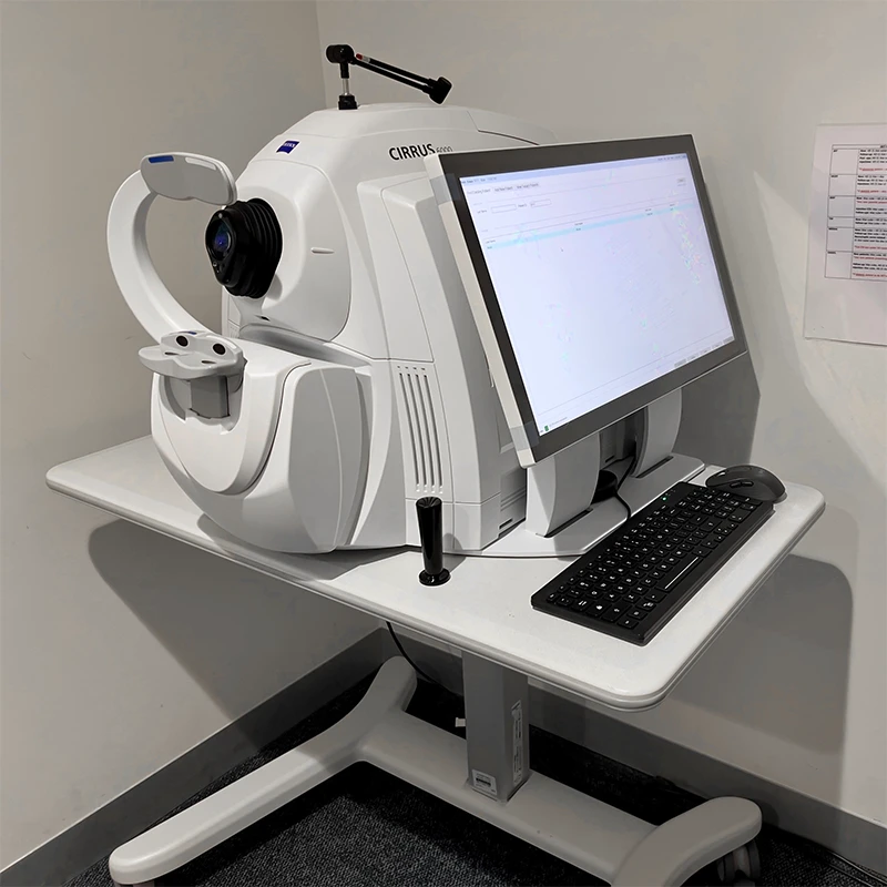
OCT with Zeiss Cirrus 6000
Uses light waves to take cross-section pictures of your retina
OCT helps in diagnosing and managing various eye conditions, including glaucoma, macular degeneration, and diabetic retinopathy.
The Zeiss Cirrus 6000 is a state-of-the-art OCT system that provides high-resolution imaging of the retina. This advanced technology allows eye care professionals to visualise the retinal structure in detail, offering critical information to assess eye health accurately.
No special preparation is required before your OCT exam. You may be asked to remove any eye makeup or contact lenses.
You will sit in front of the OCT machine and be asked to look at a target. The device will capture images of your retina without any discomfort. The scan typically takes just a few minutes.
You may resume your regular activities immediately after the scan. Your specialist will review the images and discuss the results with you during your consultation.
If you have any questions about your upcoming OCT exam, feel free to ask our staff .
Corneal Topography with Oculus Pentacam
Used to diagnose cataracts, glaucoma and corneal disease
Equipped with an automatically rotating Scheimpflug camera the Pentacam performs a complete measurement of the anterior segment of the eye in less than 2 seconds. A precise analysis of the central cornea is carried out in the process. During measurement extraneous eye movements are detected with a second pupil camera and automatically corrected during the calculation process. The software analyses and evaluates all the data acquired. These data then provide the basis for a three-dimensional model of the complete anterior eye segment.
The Pentacam analyses the entire cornea, anterior chamber and crystalline lens. This includes an objective determination of the central radii, corneal asphericity, various coloured maps of curvature and elevation, chamber angle, chamber volume and chamber elevation as well as lens transparency. Measurements are made without touching the eye and is painless.
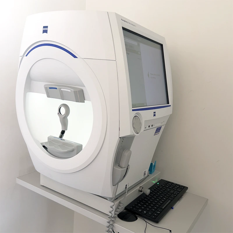
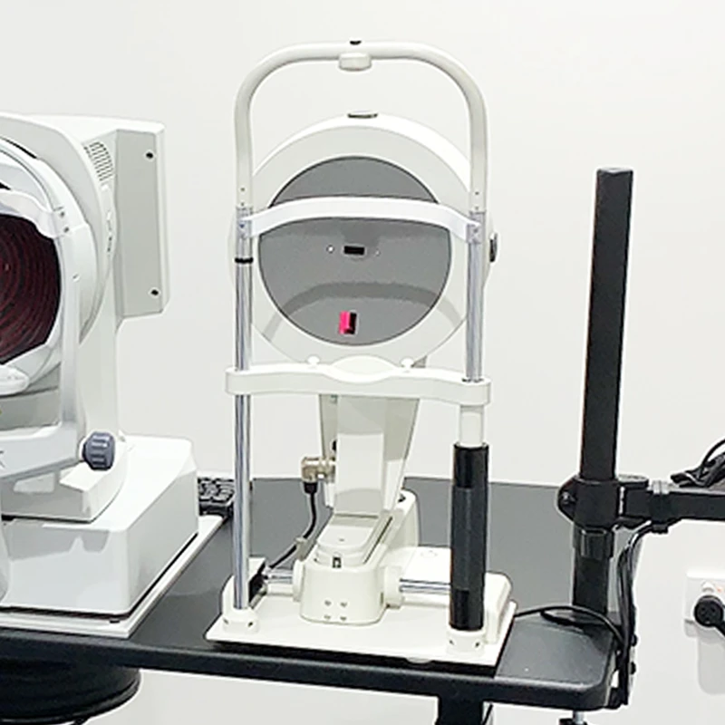
Visual Field Testing with Humphrey Field Analyser
Used to detect problems in central and peripheral vision
The Zeiss Humphrey Field Analyzer is a visual field testing device used to measure your peripheral (side) vision. It helps detect, diagnose, and monitor various eye conditions, including glaucoma, retinal diseases, and neurological disorders.
Visual field testing is essential because it provides valuable information about your overall visual function. It can help identify vision loss that may not be noticeable in everyday activities, allowing for early intervention and better management of eye conditions.
There are generally no special preparations needed before your exam. However, it’s a good idea to inform our doctors about any medications you are taking or any medical conditions.
You will sit comfortably in front of the HFA 3 device and be asked to focus on a central target. During the test, you will see flashes of light in different areas of your visual field, and you will need to press a button whenever you see the light. The test typically lasts about 5 to 10 minutes.
After the test, you can resume your normal activities immediately. Your specialist will review the results and discuss any findings or necessary follow-up appointments.
If you have any questions or concerns about your upcoming visual field exam, please don’t hesitate to reach out to our team.
For appointments and enquiries, please phone 02 9958 0552
Monday to Friday 9am - 5pm
© 2015– Northern Sydney Cataract | Privacy Policy | Disclaimer | Website by: ![]()