60 Strathallen Avenue, Northbridge NSW
Monday to Friday - 9am to 5pm
Central Retinal Vein Occlusion (CRVO)
Central Retinal Vein Occlusion (CRVO) is a sight-threatening condition that occurs when the main vein responsible for draining blood from the retina becomes blocked.
What is it?
A Central Retinal Vein Occlusion (CRVO) is a blockage of the main retinal vein that drains blood from the eye.
The blockage leads to a buildup in pressure in the vein, which causes blood and fluid to leak out onto the retina. As the central retinal veins exits the eye, it passes through a narrow opening in the lamina cribrosa that it shares with the central retinal artery. Because of this narrow opening the central vein can become compressed and lead to a blockage.
In all cases the blockage will resolve with the formation of bypass channels for blood to escape the eye. Final visual acuity is quite variable.
Risk factors may include:
- Hypertension (high blood pressure)
- Glaucoma
- Diabetes
- Hypercholesterolaemia (high cholesterol)
- Smoking
Symptoms
Symptoms for CRVO may include one or more of the following:
- Reduced vision – the loss of vision may range from partial to complete
- Black spots appearing in your vision
- Painful blind eye
Diagnosis
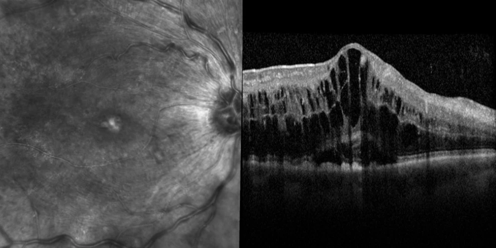
Two special tests are used to diagnose CRVO.
The diagnosis of CRVO will begin with a dilated exam of your eye. Optical Coherence Tomography (OCT) can also be used to detect macula oedema. A fluorescein angiogram may be performed to assess the severity of the interruption of retinal blood flow.
Complications
There are three types of complications that can threaten vision in BRVO.
Macula oedema
The leakage and bleeding causes the macula to swell leading to blurred vision. A prescription only medication can be injected into the eye to “dry up” this fluid.
Ischaemia
A lack of blood supply and oxygen to parts of the retina.
Neovascularisation
In severe cases of ischaemia, neovascularisation can occur where new, abnormal vessels may begin to grow. These abnormal vessels may grow throughout the eye where they can burst and bleed and cause severe visual loss. Laser (PRP) is undertaken to prevent or treat this.
Treatment
Several treatment methods are available to deal with macula oedema and to prevent neovascularisation.
Laser photocoagulation
Where retina ischaemia is prevalent, large areas of retina are treated by laser in a process known as Panretinal Photocoagulation (PRP). The aim is to prevent the abnormal vessels from growing and the resulting vision loss. Laser photocoagulation generally doesn’t improve or reduce the vision.
Intraocular injections
A type of medication called an anti-VEGF (Vascular Endothelial Growth Factor) treats both macula oedema and stops the growth of abnormal blood vessels. Repeat injections (approximately 4-6) are usually required. These usually occur approximately every 6 weeks. The thought of an injection into the eye may sound daunting but it is relatively painless and quick.
Has someone found a CRVO in your eye? At Northern Sydney Cataract, we are here to help!
If you have specific questions, please call our friendly staff on 02 9958 0552 or email us at info@nscataract.com.au
For appointments and enquiries, please phone 02 9958 0552
Monday to Friday 9am - 5pm
© 2015– Northern Sydney Cataract | Privacy Policy | Disclaimer | Website by: ![]()